vetvision.contact@gmail.com
 0789479268





KHOÁ ĐÀO TẠO ĐỌC PHIM X QUANG THÚ NHỎ: BÓNG TIM- MẠCH MÁU- PHỔI - TP. HỒ CHÍ MINH (11/08/2026) VET VISION x VSAMIA
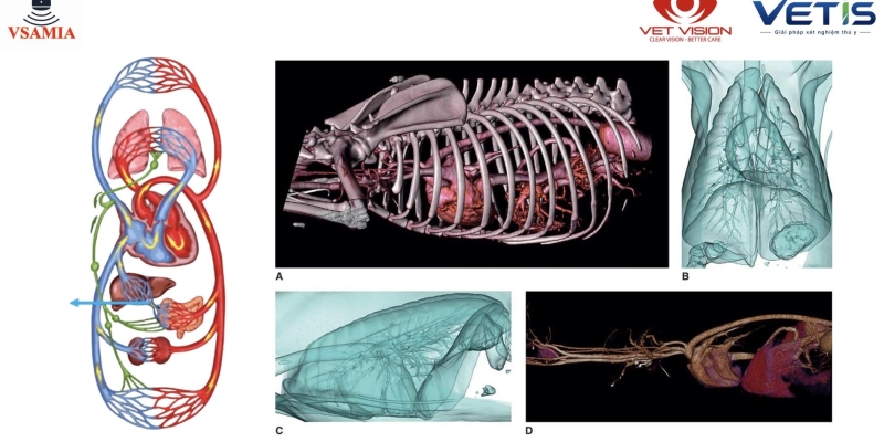
Khóa học ngắn “Đọc phim X-quang thú nhỏ: Bóng tim – Mạch máu – Phổi" do Vet Vision tổ chức ngày 11/08/2026 tập trung vào một trong những kỹ năng quan trọng trong thực hành lâm sàng thú nhỏ - đọc và phân tích phim X-quang ngực một cách có hệ thống thông qua các ca bệnh thực tế.
Thay vì chỉ ghi nhớ hình ảnh bệnh lý, khóa học hướng đến việc giúp bác sĩ xây dựng tư duy đọc phim X-quang, nhận diện các bất thường của bóng tim, hệ thống mạch máu và nhu mô phổi, đồng thời liên hệ những thay đổi trên phim với biểu hiện lâm sàng của từng bệnh thú.
Đọc phim X-quang ngực – Không chỉ nhìn hình ảnh, mà phải hiểu ý nghĩa lâm sàng
Thông qua các ca bệnh thực tế, khóa học của Vet Vision tập trung vào ba nhóm cấu trúc quan trọng:
1. Đánh giá bóng tim trên X-quang
Bác sĩ được tiếp cận phương pháp đánh giá kích thước và hình thái bóng tim, nhận diện các dấu hiệu gợi ý tim bất thường và liên hệ với những bệnh lý tim mạch thường gặp trên chó, mèo, chỉ số VHS
Việc đánh giá bóng tim không chỉ dừng ở việc xác định “tim to hay không”, mà cần xem xét hình thái, vị trí và sự thay đổi của từng vùng tim để định hướng nguyên nhân.
2. Đánh giá hệ thống mạch máu
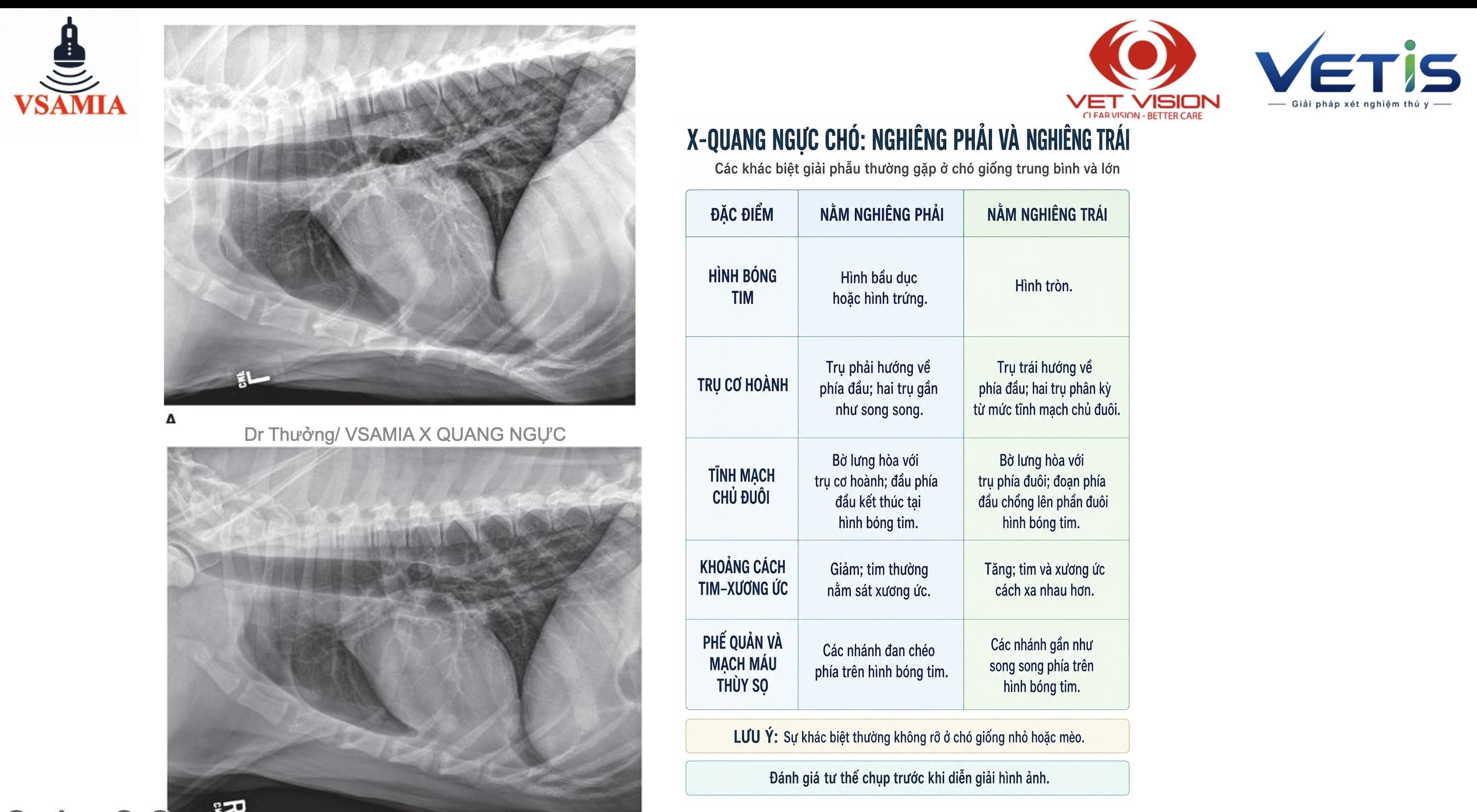
Các thay đổi về mạch máu phổi và tĩnh mạch chủ sau (caudal vena cava – CVC) có thể cung cấp những thông tin quan trọng về tình trạng tuần hoàn của bệnh thú.
Trong khóa học, bác sĩ được tiếp cận các phương pháp đánh giá định lượng như CVC/R4, qua đó hỗ trợ nhận định kích thước tĩnh mạch chủ sau và phát hiện những thay đổi có thể liên quan đến quá tải thể tích hoặc các bệnh lý tim mạch.
3. Đánh giá nhu mô phổi
Phần phổi tập trung vào cách nhận diện và phân tích các kiểu tổn thương phổi trên X-quang, từ đó giúp bác sĩ từng bước xây dựng chẩn đoán phân biệt.
Thay vì học thuộc từng hình ảnh riêng lẻ, việc phân tích theo "lung pattern"giúp bác sĩ hệ thống hóa các bất thường và liên hệ chúng với bệnh cảnh lâm sàng thực tế.
Điểm nổi bật của khóa học là nội dung được triển khai theo từng ca bệnh thực tế. Với mỗi phim X-quang, bác sĩ có cơ hội thực hành tư duy:
Thông tin bệnh thú → Đánh giá phim có hệ thống → Xác định bất thường → Định lượng khi cần thiết → Đưa ra chẩn đoán phân biệt → Liên hệ với bệnh cảnh lâm sàng.
Cách tiếp cận này giúp việc học X-quang trở nên trực quan và gần với tình huống mà bác sĩ gặp trong phòng khám, bệnh viện thú y hằng ngày.
Vet Vision- đồng hành cùng bác sĩ thú y trong đào tạo và chuyển giao công nghệ
Không chỉ là đơn vị cung cấp giải pháp chẩn đoán hình ảnh và thiết bị thú y hiện đại , Vet Vision còn luôn chú trọng đầu tư vào các hoạt động đào tạo chuyên môn nhằm góp phần nâng cao chất lượng của thú y Việt Nam.
Chúng tôi tin rằng sự kết hợp giữa kiến thức chuyên môn vững chắc và thiết bị hiện đại sẽ tạo nên nền tảng quan trọng để mang lại hiệu quả điều trị tối ưu và sự an toàn cho bệnh thú.
Vet Vision xin cám ơn các thầy và bác sĩ đã luôn đồng hành và tạo nên khóa học ý nghĩa này.


SMALL ANIMAL RADIOGRAPHIC INTERPRETATION: CARDIAC SILHOUETTE – BLOOD VESSELS – LUNGS
The short course “Small Animal Radiographic Interpretation: Cardiac Silhouette – Blood Vessels – Lungs”, organized by Vet Vision on August 11, 2026, focused on one of the important skills in small animal clinical practice – the systematic interpretation and analysis of thoracic radiographs through real-world clinical cases.
Rather than simply memorizing radiographic images of diseases, the course aimed to help veterinarians develop a systematic approach to radiographic interpretation, identify abnormalities involving the cardiac silhouette, vascular system, and pulmonary parenchyma, and correlate radiographic findings with the clinical presentation of each patient.
Thoracic Radiographic Interpretation – Not Just Looking at Images, but Understanding Their Clinical Significance
Through real-world clinical cases, the Vet Vision course focused on three important anatomical and radiographic areas:
1. Evaluation of the Cardiac Silhouette on Thoracic Radiographs
Veterinarians were introduced to methods for evaluating the size and morphology of the cardiac silhouette, identifying findings suggestive of cardiac enlargement, and correlating these findings with common cardiovascular diseases in dogs and cats, including the use of the Vertebral Heart Size (VHS).
Evaluation of the cardiac silhouette does not simply involve determining whether the heart is “enlarged or not.” It also requires assessment of the cardiac shape, position, and changes in individual cardiac contours to help determine the underlying cause.
2. Evaluation of the Vascular System
Changes in the pulmonary vasculature and caudal vena cava (CVC) can provide important information about the circulatory status of the patient.
During the course, veterinarians were introduced to quantitative assessment methods such as the CVC/R4 ratio, which can help assess the size of the caudal vena cava and identify caudal vena cava enlargement, potentially associated with volume overload or cardiovascular disease.
3. Evaluation of the Pulmonary Parenchyma
The pulmonary component of the course focused on the recognition and analysis of pulmonary patterns on thoracic radiographs, helping veterinarians gradually develop appropriate differential diagnoses.
Rather than memorizing individual radiographic images, analyzing lung patterns helps veterinarians systematically classify abnormalities and correlate them with the patient's actual clinical presentation.
Learning Through Real-World Clinical Cases
A key feature of the course was that the content was delivered through real-world clinical cases. For each radiograph, veterinarians had the opportunity to practice a systematic diagnostic approach:
Clinical information → Systematic radiographic assessment → Identification of abnormalities → Quantitative assessment when necessary → Development of differential diagnoses → Correlation with the clinical presentation.
This approach makes learning radiographic interpretation more practical and closely aligned with the situations veterinarians encounter every day in veterinary clinics and hospitals.
Vet Vision – Accompanying Veterinarians in Education and Technology Transfer
Not only a provider of modern veterinary diagnostic imaging solutions and equipment, Vet Vision also places strong emphasis on investing in professional education and training activities, contributing to the advancement of veterinary medicine in Vietnam.
We believe that the combination of solid professional knowledge and modern equipment provides an important foundation for achieving optimal treatment outcomes and ensuring patient safety.
Vet Vision sincerely thanks all the lecturers and veterinarians who have continuously accompanied us and contributed to making this course a meaningful learning experience.
Vet Vision – Accompanying veterinarians in professional education and the advancement of veterinary medicine.